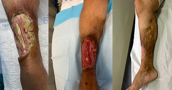
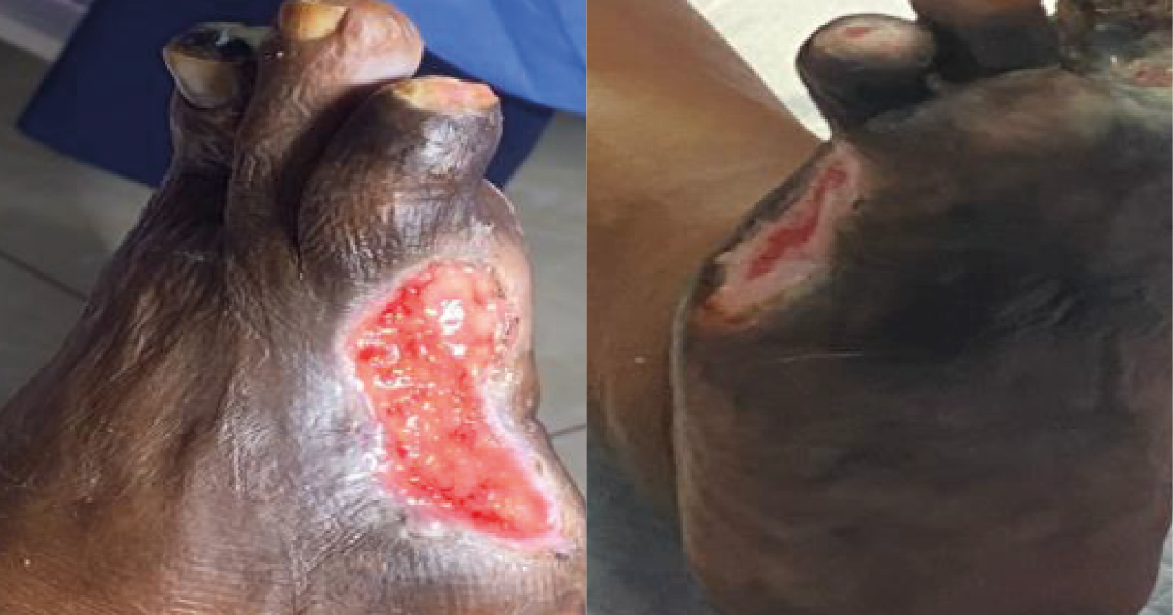
Wounds International 2(4) NovemberPractice development The diagnosis and management of necrotising fasciitis

The diagnosis and management of necrotising fasciitis

21/11/11 | Complex wounds, Infection, Skin integrity | Shady Hayek, Amir Ibrahim, Bishara Atiyeh

The diagnosis and management of necrotising fasciitisNecrotising fasciitis was first described more than a century ago, however, its diagnosis still represents a challenge for clinicians and the condition carries a high mortality rate. The management of necrotising fasciitis requires prompt diagnosis, early surgical excision and proper coordination between the multidisciplinary team to achieve the best outcome for the patient. This paper reviews the diagnostic tools used in identifying necrotising soft tissue infections as well as examining the microbiology, management and prognosis.

Introduction
Necrotising soft tissue infections (NSTIs) were first described in 1871 as 'hospital gangrene' and they continue to carry a high mortality and morbidity rate[1]. Necrotising soft tissue infections are, as the name implies, infections that affect the soft tissue by causing necrosis. They spread along the fascial layers and can be either monobacterial or multibacterial in origin.

Although they are most often associated with surgical wounds, they can also occur in tiny cracks or breaks in the skin. They are considered surgical wound infections because the treatment involves surgical excision in addition to antibiotic administration, unlike non-necrotising soft tissue infections that require antibiotics only.
However, in the years since NSTIs were first discovered, many classifications and terms have been used to describe them and there has been confusion surrounding their diagnosis and management[1-5]This review will focus on the diagnosis, management and outcome of this condition.


Microbiology
NSTI is classified into two types based on the microbial pathogens involved and the immune status of the patient[6].

Type 1
Type 1 occurs in about 20% of cases. Patients are typically healthy young individuals with an uncompromised immune system. These infections are usually monomicrobial in origin and caused mainly by group A Streptococcal infection or Staphylococcus aureus. A few cases will be caused by Clostridium species, and when associated with myonecrosis they carry a very high mortality[7]. Recently, increased cases of monomicrobial NSTI caused by methicillin-resistant Staphylococcus aureus (MRSA) have been reported[8].
Most cases of type 1 NSTI are associated with penetrating injuries or abscesses[6].

Type 2
Type 2 is the more common form of NSTI and is characterised by the presence of polymicrobial organisms of both aerobes and anaerobes. It usually occurs in people with compromised immune systems or those who are debilitated, especially those with diabetes mellitus or morbid obesity[6].
Multiple organisms have been reported including gram-positive cocci, Enterococci, Gram-positive rods, Gram-negative organisms including Klebsiella and Escherichia coli, anaerobes and even fungi[1,7,9]. The presence of such a wide spectrum of organisms means that a range of antimicrobial medication is used initially, which can be adjusted according to the culture results[1,7,9].


Diagnosis
Early diagnosis is the key to survival for patients with NSTI[1,7]. It is very important to differentiate necrotising from non-necrotising infections, as the former require surgical treatment as well as antibiotics and the latter only requires antibiotics[1]. In addition, non-necrotising soft tissue infections (like cellulitis and erysipelas) tend to occur in the skin layers themselves, whereas NSTIs tend to involve the fascial layers.

Diagnosis has been made more difficult by the different terms used to describe this condition[5], but physical examination and taking an accurate patient history remain the mainstay for establishing a diagnosis.
People who are more likely to have NSTI include those with a history of self-injecting drug misuse (although NSTIs can occur with clinician-injected medication if the sterile technique has been violated), those with diabetes, a suppressed immune system and obesity[10-12].

However, it has been repeatedly shown in larger studies that about 20% of people who develop NSTI do not have any of these precipitating factors and have an idiopathic aetiology[13-15]. It has been observed that these idiopathic cases mostly occur in people infected with group A Streptococcus[1], or MRSA[8].

Initial symptoms to look for when examining a patient with suspected NSTI include pain, swelling, erythema and tachycardia. These will progress to discolouration, blister formation, eschar formation, crepitus, swelling outside the area of skin changes and pain out of proportion to the physical findings [Fig 1][1].

Of these symptoms, the authors find a disproportionate pain level to be a crucial indicator of NSTI. It has been found, however, that although these signs and symptoms are highly specific for NSTI, their sensitivity is low as they are present in only 10-40% of patients[16, 17].

In most cases, especially those caused by monomicrobial organisms, the progression of the signs and symptoms is rapid but conversely progression can be very slow, making accurate diagnosis challenging[1].
Several diagnostic tools have been investigated that could be used in conjunction with clinical findings to help make a diagnosis as early as possible[18], including laboratory studies, imaging studies and pathological examination[1].


Laboratory studies
Wall et al[10] retrospectively studied the admission variables for necrotising and non-necrotising infections and reported that a white blood cell count greater than 15,400 x 103 cells/cc, or a serum sodium level less than 135mmol/L were associated with a necrotising infection. When combined, these two parameters had a very high negative predictive value of 99%, indicating that they are a good tool for ruling out NSTI. However, the positive predictive value was very low (26%) indicating a very low specificity.

More recently, a laboratory score was created by Wong et al[17] to differentiate between necrotising and non-necrotising infections. Called the 'Laboratory risk indicator for necrotising fasciitis score' (LRINEC), it identified six independent laboratory variables associated with NSTI and gave each one of them a number of points [Table 1].

The summation of the points gives a score that ranges between 0-13. Three groups were then identified according to their risk for NSTI:

  • If the score is 5 or less, then the probability of NSTI is low (less than 50%)
  • If the score is 6 or 7, then the probability of NSTI is 50-75%
  • If the score is 8 or more, then the probability of NSTI is very high (more than 75%).


This tool was found to have a high positive predictive value (92%) and also a high negative predictive value (96%) for intermediate and high-risk groups[1, 11].

Another test, the Acute Physiology, Age and Chronic Health Evaluation (APACHE II) score has been found to be non-specific for NSTI diagnosis, but helpful in predicting the prognosis for people with the infection[9, 19]. Yilmazlar et al[19] reported that patients who had an APACHE II score greater than 13 had an 86% mortality rate. All patients who died during the study had an APACHE II score of greater than 20.

All of these scores and laboratory parameters are supposed to aid in diagnosis, but it is important to remember that none of them replace the physical examination and assessment of an expert physician in making an early and accurate diagnosis[9].